P
p.a.- oder a.p.-Projektion? (Text)
PAH (Liste der Berufskrankheiten) (Text)
Pancoast-Tumor (Text)
(Bild)
-, MR (Text)
Pankreasmetastase (Bild)
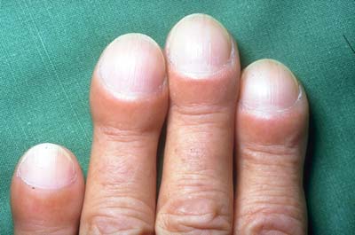
Paraneoplasie bei BC (Bild)
-, Uhrglasnägel (Text)
(Bild)
-, Periostverdickung
(Bild)
Paraquat (Text)
(Text)
Parathion (Text)
Parenchymbänder (Text)
(Def.)
Parenchymfibrose nach
Beatmung (Text)
Parenchymverdichtung
(Text)
(Def.)
Parkotil® (Text)
Patienten-Aufklärung (Text)
PcP (Pneumocystis carinii)
(Bild)
- und Tb (Bild)
-, Rö-Zeichen (Text)
-, Verlauf (Bild)
-, vorzeitiges Emphysem (Bild)
Penicillamin
(Text)
Penicilline (Text)
(Text)
Perfusionsdefekte, szintigraphische
(Bild)
Pergolidmesilat (Text)
Perikard
(Bild)(Bild)
-
dicke
(Normalwerte)
- drainage
(Bild)
- emphysem (Rö.-Z.)
- erguß (Bild)
- falte, DD Lymphom (Bild)
- krankheiten (Text)
-, luxatio cordis (Bild)
- umschlagfalte (Text)
- verkalkung (Bild)
zyste (Bild)(Bild)
(Rö.-Z.)
perikardiales Fett
(Bild)
Perikarditis (Text)
Periostverdickung
(Bild)
peripher
(Def.)
Pest (Text)
Pflanzenschutzmittel (Text)
Phenytoin (Text)
Phosgen (Text)
Pièrre-Marie-Bamberger (Bild)
Pilzinfektion bei AIDS, Rö-Zeichen
(Text)
Pilzpneumonie (Bild)
(Text)
Plasmozytom (Bild)
Plattenatelektase (Text)
Plattenepithelkarzinom (Bild)
(Makropathologie)
Pleura (Text)
-, gespaltene (split pleura sign) (Text)
-, KM-Bolus (Text)
-, Metastasen (Text)
-, Tumorinfiltration (Text)
- parietalis (Text)
- viszeralis (Text)
Pleuraasbestose (Bild)
(Bild)
(Text)
Pleuraempyem, Formanalyse (Text)
(Bild)
- -, DD Abszeß, Rö-Zeichen
(Text)
Pleuraerguß,
- -, abgekapselter (Text)
- -, DD (Text)
- -, hämorrhagischer (Text)
- - im HRCT, DD (Tabelle)
- -, Liegeaufnahme (Text)
- -, massiver, Rö-Zeichen (Text)
- -, Rö-Zeichen (Text)(Text)
- - und Herzinsuffizienz, Rö-Zeichen
(Text)
- -, Ursachen (Text)
- -, sonographische Zeichen (Text)
- -, subpulmonaler (Text,
Bild)
Pleurafinger (Bild)
- infiltration (Bild)
- karzinose (Bild)
Pleurakuppenschwielen (Text)
pleurale Raumforderungen
(Bild)
pleurale Umschlagfalte (Text)
Pleuralipom (Text)
Pleuramesotheliom
-, bei Asb.(Bild)
(Bild)
Makroanatomischer
Schnitt
-, Erscheinungsformen (Bild)
-, Lungenmetastasen (Bild)
-, Rö-Zeichen (Text)
pleuraparallele Linien (CT) (Text)
(Bild)
Pleuraplaques (Bild)
(Bild:
Makropathologie
(Text)
- schwielen, (Bild),
Rö-Zeichen (Text)
- tumor, Malignitätskriterien (Text)
- -, Rö-Zeichen (Text)
- tumor, fibröser (Text)
- -, bösartiger (Text)
|

|
Pleuraverdickung
(Text)
- bei Asbestose (Bild)
(Text)
(DD)
- bei Asbestose, DD (Bild)
-, Dignitätskriterien (Text)
-, flächige bei Asbestose (Bild)
Pleuraverkalkung (Text)
(DD)
- verletzungen (Text)
Pleuritis carcinomatosa (Bild)
Pleuritis exsudativa (Text)
Pleuritis exsudativa tuberculosa (Bild)
Pneumatozele (Text)
(Text)
(Def.)
-, Entwicklung (Bild)
-, bei
AIDS
(HRCT,
Text)
Pneumektomie,
postop. Verlauf (Bild)
(Bild)
Pneumocystis
carinii Pneumonie: siehe auch PcP
(Text)
- im HRCT, DD (Tabelle)
Pneumogramm (Bild)
Pneumokoniosen (Text)
Pneumonektomie, (Bild)
Rö-Zeichen (Text)
Pneumomediastinum (Text)
(Bild)
Pneumonie
-, akute interstitielle, Rö-Zeichen
(Text)
-, alveoläre (Text)
-, ambulant erworbene, (Text)
(Rö-Z.)
-, -, Erregerspektrum (Text)
-, atypische Erreger (Text)
-, auf Liegeaufnahme, Rö-Zeichen
(Text)
-, bronchoalveoläre (Text)
-, chronische (Text)
-, diffus, beidseitig (Text)
-, Erreger bei nosokomialer (Text)
-, Erregerzuordnung (Text)
-, homogene Verdichtungen (Text)
-, idiopathische interstitielle (Text)
-, immungeschwächter Patienten
(Text)
-, interstitielle (Bild)
(Rö.-Z.)
-, Klassifikation (Text)
-, Komplikationen (Text)
-, lobäre (Rö.-Z.)
-, mit Bronchogramm (Bild)
-, nosokomiale (Text)
(Text)
-, rezidivierende (Text)
-, Verlauf (Text)
Pneumonitis, radiogene, Rö-Zeichen
(Text)
-, -, aktive (Bild)
Pneumoperikard
(Bild)
(Rö.-Z.)
Pneumoseroperikard
(Bild)
Pneumothorax (Bild)
(Text)
- auf Liegeaufnahmen (Text)
(Bild)
(Rö-Z.)
-, Fehldiagnose (Bild)
-, Liegethorax (Text)
-, partieller
(Bild)
-, Rö-Zeichen (Text)
-, traumatischer (Bild)
polygonales Muster (Text)
Polygone im HRCT, DD (Tabelle)
polyzystische Organdegeneration (Bild)
Portkathetersystem (Bild)
Positionierung (Thoraxaufnahme) (Text)
postoperative Veränderungen, Rö-Zeichen
(Text)
Postpneumektomie-Syndrom (Bild)
Postprimär-Tb (Bild)
(Rö-Z.)
(Text)
Posttransplantationslymphom (Text)
(Rö.-Z.)
prätracheal-retrocavalen Lymphknoten
(Bild)
Primärkomplex
(Bild)
(Text)
Rö.-Z. (Text)
Primärtuberkulose (Text)
(Bild,
Sammlung)
Primärtumorstaging (Text)
Procainamid
(Text)
produktive Reaktion (Text)
produktive Herde der Tb (Bild)
(
Text)
Pseudoläsionen, szintigrafische
(Bild)
Pseudolobus venae azygos (Text)
Pseudoplaque (Text)
(Def.)
(Bild)
Pseudotumor (vanishing tumor) (Bild)
- bei Erguß (Text)
Psittakose (Bild)
Pulmonalarterien (Bild)
- Aneurysma (Bild)
- Fehlbildungen (Bild)
- rechts, Weite (Normalwerte)
pulmonale Hypertonie (Text)
-, WHO-Klassifikation 1998 (Text)
Pulmonalgefäße (Bild)
Pulmonalisangiographie
(Bild)
Pulmonalishauptstammweite (CT) (Text)
(Normalwerte)
Pulmonaliskatheter (Text)
Pulmonalisklappe (Bild)
Pulmonalisweite (Text)
Punktion, transthorakale (Bild)
- (Text)
|